Our Services
MRI Images
Every patient immediately receives a complimentary CD of their images to utilize as a reference
for their personal health history and to help provide continuity of care to other health care professionals.
Brain MRI
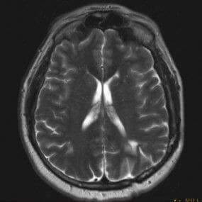
Brain imaging can look at the brain for evaluation of tumors, aneurysms, bleeding in the brain, nerve injury, and other problems, such as damage caused by a stroke. A Brain MRI can also find problems of the orbits (eyes and optic nerves), internal auditory canals (ears and auditory nerves), and pituitary glands.
Orbit MRI
The facial orbit is the cavity or socket of the skull in which the eye and its appendages are situated. Orbital Imaging is used to discover tumors, infection, chronic diseases, and optic neuropathy. Other conditions evaluated with orbital imaging include papilledema (swelling head of the optic nerve), enlarged eye muscles (seen in thyroid conditions), infection or tumors of the lacrimal gland, and enlargement of vessels that supply and drain the eye area.
Adrenal Glands MRI
Adrenal glands protocol is an MRI protocol comprising a group of MRI sequences put together to further assess indeterminate adrenal lesions, in particular, lipid-poor adenomas. In addition, an Adrenal Gland MRI scan may be done to check for certain cancers or other illness.
Pituitary Gland MRI
The pituitary gland is a pea sized structure in the middle of your brain just behind your eyes. It sits inside a bone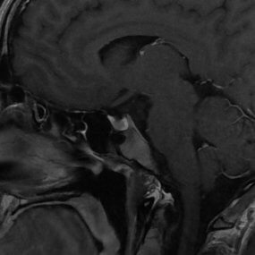 cavity called the sella dorsica. The gland is attached to the brain by a thin stalk called the infundibulum. Pituitary gland MRIs are used to discover tumors of the pituitary gland, such as microadenomas, craniopharyngiomas, apoplexy tumors, and cysts.
Internal Auditory Canal
This is a large nerve that conducts hearing and balance signals from your inner ear structures. This nerve can become damaged, inflamed or give rise to a tumor commonly called an “acoustic neuroma.” Most Internal Auditory Canal MRIs are ordered to rule out an acoustic neuroma in patients who are experiencing dizziness, ringing in the ear or hearing loss.
Magnetic Resonance Angiography
MRA or Magnetic Resonance Angiography is a type of magnetic resonance image (MRI) scan. MRI scans are used to look at blood vessels, and the flow of blood through them is called magnetic resonance angiography (MRA). An MRA evaluates blood vessels. The blood vessels in the neck (carotid and vertebral arteries) and brain are frequently studied by MRA to look for areas of narrowing or dilatation. In the abdomen, the arteries supplying blood to the kidneys are also frequently examined with this technique. The extremities (arms, legs) can also be studied for narrowing. MRA scans can find problems of the arteries and veins, such as an aneurysm, a blocked blood vessel, or the torn lining of a blood vessel (dissection). Sometimes contrast material is used to see the blood vessels more clearly. Like an MRI, magnetic resonance angiograms (MRA) use a magnetic field and pulses of radio wave energy to make pictures of blood vessels inside the body.
Kidney MRI
Kidney MRI It can also provide a functional assessment of your kidneys, including information about their GFR, blood volume and perfusion, diffusion, and oxygenation. Your healthcare team will use these images to detect possible issues with your kidney health. MRI scans of your kidneys can help see conditions like kidney cancer, chronic kidney disease, renal vein thrombosis, and the presence of tumors, masses, stones, or infection.
Musculoskeletal (MSK)
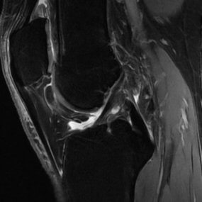
Musculoskeletal imaging can evaluate virtually all of the bones and joints, derangement, infection, inflammation, post-trauma, and tumors. Soft tissue evaluation includes tendons, ligaments, muscles, cartilage, and bone injuries. MRI can help diagnose various mass compositions and vascular pathologies.
Pelvic MRI
Pelvic imaging investigates pathology and detection of a local invasion of rectal vascular pathologies, assessing the anatomy in peri-anal fistulas, infection, and inflammation. For women, pelvic MRI is used to evaluate the ovaries and uterus as a follow-up to an ultrasound exam which showed an abnormality, or cervical carcinomas, and to evaluate endometrial cancer. For men, pelvic MRI is sometimes used to evaluate prostate cancer.
Brachial Plexus MRI
The brachial plexus represents a complex network of nerves formed from the ventral rami of the lower cervical nerves (C5-C8) and the greater portion of the ventral ramus of the first thoracic nerve (T1). The ventral rami or roots 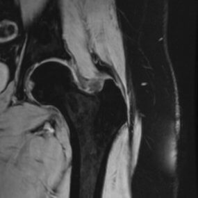 coalesce to form trunks, divisions, cords, and branches, which provide motor and sensory innervation to the upper limbs and thorax. Brachial plexus imaging allows for visualization of a wide range of pathologies such as birth trauma, high-speed trauma, neoplasms (intrinsic or extrinsic masses) or post-radiation injury.
Abdominal MRI
Abdominal imaging is most frequently used to further evaluate an abnormality seen on another test, such as an Ultrasound (US) or Computed Tomography (CT) scan. The MRI exam is usually tailored to look at specific organs or tissues, such as the liver, adrenal glands, kidneys or pancreas, in addition to various tumors, congenital abnormalities, and metabolic disorders.
Magnetic Resonance Cholangiopancreatography
Magnetic resonance cholangiopancreatography (MRCP) is a special type of magnetic resonance imaging (MRI) exam that produces detailed images of the hepatobiliary and pancreatic systems, including the liver, gallbladder, bile ducts, pancreas, and pancreatic duct. This is used to help evaluate tumors, stones, inflammation or infection, and to 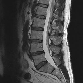 evaluate patients with pancreatitis to detect the underlying cause.
Spine MRI
Spine imaging includes the anatomy of the cervical, thoracic, lumbar, and lumbosacral region. MRI scans are most commonly used to evaluation herniated discs or narrowing of the spinal canal (spinal stenosis) in patients with neck, arm, back and/or leg pain. It is also the imaging tool to look for a recurrent disc herniation in patients with a history of back surgery.